Detection of Multidrug-Resistant Acinetobacter junii in a Himalayan Fresh water Ecosystem: A Pilot Study from Kumaon Kosi River
Manohar Kumar
 , Shivangi Singh
, Maneet Kumar Chakrawarti
, Bavya Krishna
, Pavithra B
, Kasturi Mukhopadhyay
and Madhuri Singh
*
, Shivangi Singh
, Maneet Kumar Chakrawarti
, Bavya Krishna
, Pavithra B
, Kasturi Mukhopadhyay
and Madhuri Singh
*
1
Environmental Microbiology and Antimicrobial Research Laboratory,
School of Environmental Sciences,
Jawaharlal Nehru University,
New Delhi,
India
http://dx.doi.org/10.12944/CWE.21.1.8
Copy the following to cite this article:
Kumar M, Singh S, Chakrawarti M. K, Krishna B, Pavithra B, Mukhopadhyay K, Singh M. Detection of Multidrug-Resistant Acinetobacter junii in a Himalayan Fresh water Ecosystem: A Pilot Study from Kumaon Kosi River. Curr World Environ 2026;21(1). DOI:http://dx.doi.org/10.12944/CWE.21.1.8
Copy the following to cite this URL:
Kumar M, Singh S, Chakrawarti M. K, Krishna B, Pavithra B, Mukhopadhyay K, Singh M. Detection of Multidrug-Resistant Acinetobacter junii in a Himalayan Fresh water Ecosystem: A Pilot Study from Kumaon Kosi River. Curr World Environ 2026;21(1).
Download article (pdf)
Citation Manager
Publish History

Introduction
The presence of residual antibiotic load within aquatic ecosystem has led to development of antimicrobial resistant bacteria (ARBs) which in turn exacerbates the global public health issue of antimicrobial resistance (AMR).1Recognized by the World Health Organization (WHO), AMR is one of the most prevailing global health issues and jeopardizes the effective treatment of bacterial infections, contributing to increased mortality, morbidity and cost of healthcare worldwide.2 In 2019, approximately 4.95 million death and 1.27 million infections associated with antimicrobial resistance (AMR) was reported globally.3Gram-negative bacteria, particularly those belonging to the Acinetobacter genus, are among the most concerning contributors to this burden due to their ability to persist in diverse environments and rapidly acquire resistance determinants.4Acinetobacter species are Gram-negative, aerobic, non-motile coccobacilli commonly found in soil, freshwater, and hospital environments.5,6Although traditionally considered low-virulence organisms, several species have emerged as opportunistic pathogens, primarily affecting immunocompromised patients and individuals in intensive care settings.7Their pathogenicity is largely driven by immune evasion strategies such as capsular polysaccharide production and lipopolysaccharide (LPS)-mediated stimulation of Toll-like receptor 4 (TLR4), leading to systemic inflammation and septic shock. 8,9The resistance mechanisms employed by Acinetobacter spp., are multifactorial, including the production of B-lactamases, reduced outer membrane permeability, active efflux pumps, target site modifications, and robust biofilm formation.10,11
Among these species, Acinetobacter baumannii has received the most clinical attention as a major cause of nosocomial infections (HAIs), particularly in ICUs.7,12 However, less-studied species such as Acinetobacter junii are now emerging as important players in both clinical and environmental AMR contexts. A. junii is one of over 70 recognized species within the Acinetobacter genus.7 Historically under-recognized, recent reports have linked A. junii to a variety of opportunistic infections, including neonatal septicaemia, bacteraemia in cancer patients,13-15 urinary tract infections, and ocular infections such as corneal ulcers and perforation.16, 17 Of growing concern is its ability to harbour and disseminate resistance genes such as NDM-1, OXA-58, and IMP-type carbapenemase that get transferred from one species to another via horizontal gene transfer (HGT).18Environmental isolates, particularly from poultry and wastewater sources, have demonstrated resistance to clinically significant antibiotics, suggesting an ecological bridge between environmental, animal, and human health sectors.5, 18
Large-scale genomic studies have reported limited genetic divergence between clinical and environmental A. junii isolates, indicating possible cross-sector transmission routes.19, 20Despite this, A. junii remains underrepresented in research, with fewer than 170 scientific publications indexed in NCBI as of August 2023.19 This under-characterization poses a risk, particularly in the context of environmental AMR surveillance and early detection of emerging threats.19 Moreover, naturalwater bodies, especially those perceived as pristine, may also serve as silent reservoirs for transmission of antibiotic-resistant bacteria, necessitating their inclusion in AMR surveillance networks.In this context, the present study investigates the infiltration of multidrug-resistant A. junii into a relatively unpolluted freshwater ecosystem the Kosi River in Uttarakhand, India. Through systematic isolation, biochemical and molecular identification, and antibiotic susceptibility testing, we highlight the environmental dimension of AMR in environmental isolates of A. junii.
Material and Methods
Study Area
Water samples were collected from the Kosi River in Nainital (Lohali village) Uttarakhand (29°30'02.5"N, 79°30'17.0"E) on 23 June 2023, as depicted in Figure 1. The Kosi River originates from Dharapani Dhar near Kausani in the Almora district of the Kumaon region and serves as an important tributary of the Ganga River system. Spanning approximately 116.14 km in length, the river plays a vital role in irrigation, hydropower generation, and public water supply. Additionally, it supports rich fish biodiversity and contributes to the local economy through activities such as fishing, sport fishing, and water-tourism.1, 21
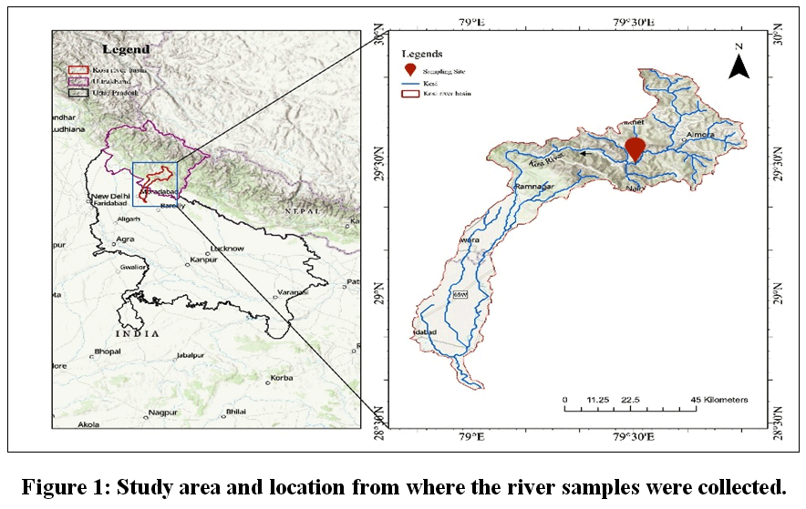 | Figure 1: Study area and location from where the river samples were collected.
|
Isolation,Identification, Biochemical and Molecular Characterization ofBacterial Isolates
The samples were collected,serially diluted and plated onnutrient rich agar like Brain-Heart Infusion (BHI)as well as selective and differential media like Mannitol Salt Agar (MSA) and Baird-Parker Agar (BPA). The oxacillin susceptibility test involved inoculating bacteria on Brain-Heart Infusion (BHI) supplemented with oxacillin (1µg/mL); bacterial growth indicates resistance to oxacillin and thus a positive result for resistance.22 To identify and characterize the bacterial isolates, a series of biochemical tests like catalase, coagulase, oxidase and Gram stainingwere performed. The catalase test was done to check the production of bubbles indicating a positive result due to the release of oxygen when exposed to hydrogen peroxide (H2O2). The coagulase test was performedto detect free coagulase secreted by bacteria,formation of a clot indicates a positive result.23 The oxidase disc was used to check the cytochrome c oxidase activity which turn the disc blue giving positive result.24 Finally Gram staining was done to check the morphology of each isolates in order to classify it as Gram positive or Gram negative bacteria.25
For molecular identification, genomic DNA was extracted from purified colonies, further the quality of DNA was checked on NanoDrop 2000 Spectrophotometer (Thermo Scientific) and 16S rRNA gene amplification was done via Polymerase Chain Reaction (PCR) using universal primersand sequencing were conducted. The bacterialsequences were later identified through NCBI database using BLAST to determine the closest phylogenetic affiliations.
Antibiotic Susceptibility Test (AST)
Kirby-Bauer disk diffusion method was used to test antibiotic susceptibility profile byfollowing Clinical & Laboratory Standards Institute (CLSI 2024) guidelines. All Acinetobacter junii isolates and the control strain (Acinetobacterbaumannii)weretested against a panel of antibiotics: amikacin, ampicillin, cefepime, cefuroxime, ceftazidime, ciprofloxacin, cefoxitin, gentamicin, doripenem, meropenem, imipenem, streptomycin and tigecycline. The inhibition zones were measured and compared with CLSI guidelines to classify them as sensitive, intermediate, or resistantisolates.
Minimum Inhibitory Concentration (MIC) Assay
The minimum inhibitory concentration (MIC)was determined for polymyxin B and colistin using broth microdilution methods, given their relevance as last-resort antibiotics against Gram-negative multidrug-resistant bacteria as per CLSI guidelines.25 Selected environmental A. junii isolates, GRS 3 and GRS 5 and control strain A. baumannii were studied. In brief, a sterile 96-well microtiter plate was prepared with two-fold serial dilutions of the antibiotic in Mueller Hinton II Broth Cation adjusted(MHB)across columns 1 to 10, concentration ranging from 256 µg/ml to 0.5 µg/ml. Column 11 served as a positive growth control (media + bacteria), and column 12 as a negative control (media only).
Next, bacterial cultures were adjusted to ODooo of 0.3–0.4 (~108 CFU/ml) and diluted to 106 CFU/ml. Then, 50 µl of this suspension was added to wells from columns 1–11 containing antibiotic. The plate was incubated at 37°C without shaking for overnight growth. Bacterial growth was assessed the next day in each column based on visible turbidity.
Results
Isolation and Identification of Bacterial Isolates from Kosi River Water Sample
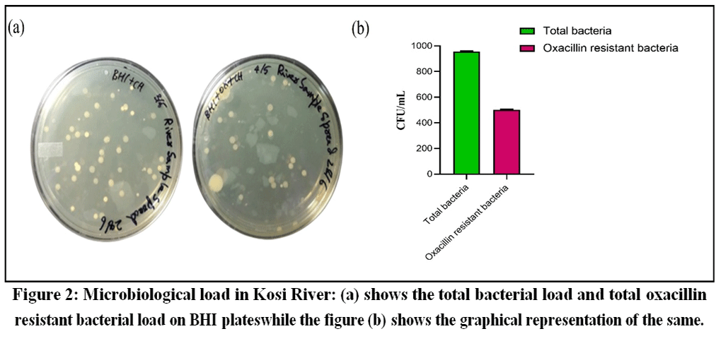
The collected water sample was brought to the laboratory and 50 µL of it was spread on BHI Agar with and without 1µg/ml oxacillin for total bacterial load and presumptive oxacillin resistant bacteria respectively. After an overnight incubation at 37°C it was observed that the BHI media without oxacillin had 48 colonies and the BHI media containing oxacillin had 25 colonies. Therefore, the total load of bacteria in Kosi River sample was 960 CFU/mL, and oxacillin resistant bacterial load was 500 CFU/mL as shown in Figure 2 (a and b). Further, glycerol stocks were prepared from the primary culture of these bacterial isolates and stored at -80°C for future use. The isolation from the Kosi River water samplesindicated a substantial bacterial load (960CFU/mL) reflecting active microbial presence, possibly due to anthropogenic influences.26The Oxacillin supplemented BHI plates gave 500 CFU/mL count, highlighting 50% of total culturable bacterial population showing presumptive resistant to B-lactam antibiotics. This finding raises concern regarding the dissemination of antibiotic resistance in freshwater sources, suggesting discharge of untreated sewages from hospital, domestic and agricultural runoff.27
 |
|
Simultaneously, the isolates with different morphology were selectively isolated from oxacillin supplemented BHI plates and restreaked multiple times for purification. After purification the isolates were restreaked on MSA, and BPA plates. On MSA, none of the 25 isolates demonstrated growth, indicating a negative result and suggesting the absence of Staphylococcus spp. Similarly, no growth on BPA was observed,whereas only GRS13, which developed grey-black colonies, while the remaining isolates showed no growth, ruling out presumptive Staphylococcus identification for most of the isolatesas shown in Figure 3.
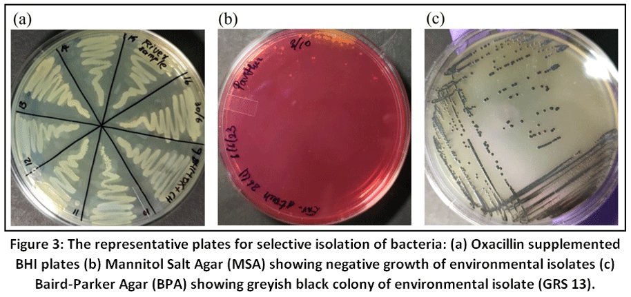 | Figure 3: The representative plates for selective isolation of bacteria: (a) Oxacillin supplemented BHI plates (b) Mannitol Salt Agar (MSA) showing negative growth of environmental isolates (c) Baird-Parker Agar (BPA) showing greyish black colony of environmental isolate (GRS 13).
|
Subsequently, biochemical tests like catalase, coagulase, oxidase and Gram-staining tests were performed to classify the isolated strains.Out of 25, only 24 isolates were catalase positive, coagulase negative, oxidase negative with no blue colouration on disc and Gram stainingrevealed the coccobacillus shape suggesting the isolates to be Gram-negative bacteria as shown in Figure4 respectively.
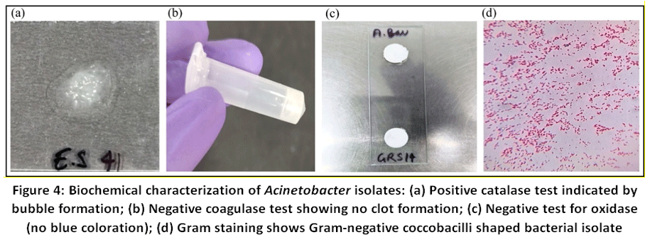 | Figure 4: Biochemical characterization of Acinetobacter isolates: (a) Positive catalase test indicated by bubble formation; (b) Negative coagulase test showing no clot formation; (c) Negative test for oxidase (no blue coloration); (d) Gram staining shows Gram-negative coccobacilli shaped bacterial isolate
|
ll the 24 presumptive isolates were identified using 16s rRNA gene sequencing and it confirmed to be A. juniiwhich were carried forward for subsequent analysis. This predominant presence of A. juniiin river system, warrants further investigation into its environmental reservoirs and transmission pathways.
Antibiotic Resistance Profiles of A. Junii Isolates from Kosi River
All twenty-four Acinetobacter isolates were subjected to the Kirby-Bauer disc diffusion susceptibility test for amikacin, ampicillin, cefepime, cefuroxime, ceftazidime, ciprofloxacin, cefoxitin, doripenem, gentamicin, imipenem, meropenem, streptomycin and tigecycline. The control Acinetobacter baumannii, is an MDR strain which we used as a control strain for Kirby-Bauer Test. The antibiotic susceptibility profile of all environmental isolates reveals predominantly moderate to high sensitivity for all the antibiotics. While, the reference strain A. baumannii showed multi-drug resistance phenotype, especially against beta-lactams class of antibiotics. All isolates were shown to be resistant to cefepime, the isolate (GRS5) (Figure 5) and isolate (GRS3) exhibited resistance and intermediate resistance to ceftazidime, whereas, 8 isolates (GRS3, GRS5, GRS18, GRS19, GRS20, GRS21, GRS22 and GRS24) showed intermediate resistance to cefoxitin (2nd generation cephalosporins) (Table 1). Compared to MDR phenotype of Acinetobacter baumannii, all the A. juniishowed higher susceptibility towards most antibiotics primarily due to isolation from pristine environment.
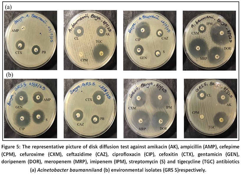 | Figure 5: The representative picture of disk diffusion test against amikacin (AK), ampicillin (AMP), cefepime (CPM), cefuroxime (CXM), ceftazidime (CAZ), ciprofloxacin (CIP), cefoxitin (CTX), gentamicin (GEN), doripenem (DOR), meropenem (MRP), imipenem (IPM), streptomycin (S) and tigecycline (TGC) Antibiotics (a) Acinetobacter baumanniiand (b) environmental isolates (GRS 5)respectively.
|
The intermediate resistance to cefoxitin by 8 isolates also points towards the possible development of resistance in process. These findings reinforce aquatic environmentas a potential reservoir of AMR particularly where animal and human interface overlap posing serious concerns for public health. Nonetheless, the antibiotic susceptibility test using disc diffusion test revealed an alarming trend: all A. junii isolates were found to be resistant to cefepime, a fourth-generation cephalosporin commonly used in clinical settings. Although bacterial isolates showed intermediate resistance to ceftazidime, cefoxitin and resistance to cefepime, suggesting either a B-lactamaseresistance especially the Extended Spectrum Beta-lactamase (ESBL) and AmpC activity. Overall, the environmental isolates showed higher susceptibility to tigecycline and carbapenems indicate the retained activity of carbapenemase activity, highlighting their continued efficacy as last-resort drug. The percentage of antibiotic susceptibility of A. junii isolates showing high level of susceptibility (100%)against ampicillin, ciprofloxacin, gentamicin, streptomycin, doripenem, imipenem, meropenem, cefuroxime and tigecycline as shown in Figure 6 and Table 1.
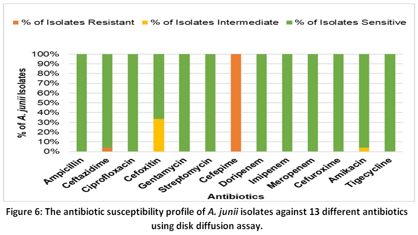 | Figure 6: The antibiotic susceptibility profile of A. junii isolates against 13 different antibiotics using disk diffusion assay.
|
Further the MIC values of selected isolates that were multi-drug resistant was determined against polymixin B and colistin. The MIC is defined as the minimum concentration of antibiotic at which no bacterial growth is visible, indicating complete inhibition of bacterial proliferation, as mentioned in Table 1. The MIC assay results provide a quatitative assessment of antibiotic resistance against polymyxin B and colistin in GRS 3 and GRS 5particularly important in treatment of Gram-negative infections, helping us assess the antibiotic selective pressure on environmental isolates. The MIC assay revealed the isolate GRS5 to have relatively higher MIC value (<2µg/ml) as compared to GRS3 (<0.5) but still susceptible to the polymixin B and colistin suggesting the retained efficacy of last-resort drugs. Nonetheless, the colistin and polymixin B remains effective against environemntal isolates but routine surveillance of antibiotic susceptibility patterns in environmental settings for AMR of both pathogenic and non-pathogenic isolate is needed.
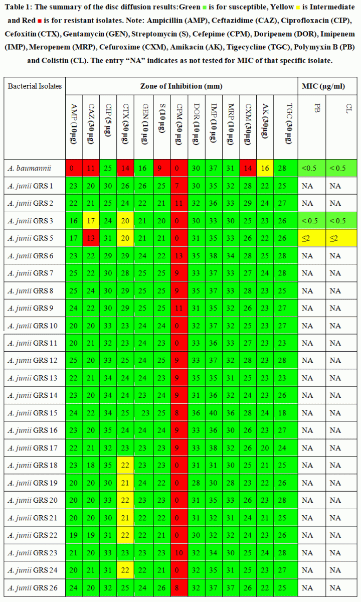 | Table 1: The summary of the disc diffusion results: Green is for susceptible, Yellow is Intermediate and Red is for resistant isolates.
|
Discussion
Antimicrobial resistance (AMR) is the ability of microbe to resist the antibiotic and survive in their presence.28AMR has become a global public health crisis now due to constant cycling of AMR bacteria between human, animals and environment.29 Earlier it was reported mostly from clinical samples and from clinical settings only.25However, the presence of antimicrobial resistant bacteria and antibiotic resistance related genetic determinants are ubiquitously present in environmental settings including air, water and soil samples due to contamination of these samples with residual amount of antibiotics allowing bacteria to evolve with molecular mechanisms of AMR.30
This study focuses on analyzing the total bacterial load and oxacillin resistant bacterial load. We found 960 and 500 CFU/mL of total bacterial load and total oxacillin resistant bacterial load respectively in the Kosi River water samples. All the 24 bacterial isolates growing on the oxacillin plate were identified as A. junii in the river ecosystem using 16S rRNA gene sequencing. Further, multidrug resistance (MDR) profiling was performed on the A. juniiisolates, this analysis provides insights into the resistance potential and associated public health risks.The isolates in this study confirmsthe presence of Gram-negative bacteria, even in pristine water bodies which were 100% resistant to cefepime (a 4th generation cephalosporin) while 8 isolates (33%) were intermediate resistant to cefoxitin (a 2nd generation cephalosporin). This may be due to extensive use of the latest drug i.e., cefepime against Gram-negative bacteria as compared to cefoxitin due to reduced usage of conventional antibiotics rendering less selective pressure on bacteria to evolve resistance.Notably, isolates GRS 3 and GRS 5 exhibited multidrug resistance against cephalosporin class of antibiotics, which is particularly worrying from a public health perspective.Additionally, the emergence of intermediate resistanceagainst polymyxin B and colistin (the last-resort antibiotics against Gram-negative bacteria)further underscores the need to perform such monitoring studies to estimate the AMR burden in environmental ecosystem. While extensive research has focused on A. baumannii, comparatively limited work has been conducted on A. junii.31 Even though A. junii is considered less pathogenic than A. baumannii, its ability to harbour AMR determinants makes it an emerging threat that cannot be overlooked.32
This study is limited by random sampling and restricted sampling locations. To obtain a clearer and more representative picture of AMR dissemination in the Kosi River, wider surveillance across the entire river stretch is required, along with long-term sampling at multiple time intervals. Additionally, molecular investigations are essential to bridge the current knowledge gap and to better understand the genetic basis of resistance and potential transmission pathways. The co-selective pressure of heavy metals is precursor to spreading and persistence of ARGsin the environmental settings due to the genetic involvement of metal resistance genes toARGs. Antimicrobial resistance is being accelerated by co-resistance, cross-resistance, andco-regulation in addition to horizontal gene transfer and mobile genetic elements.33 Forinstance, previously Talat, et al., study shows heavy metal resistance genes such as meroperon and CopR were cooccurring with many ARGs alongside hospital waste water suggest co-selection, in which metal contamination may indirectly promote antibiotic resistance.34Despite these limitations, the detection of AMR-carrying A. junii suggests that the river may serve as an AMR reservoir, with the potential to contaminate connected water bodies and contribute to the spread of resistance on a larger scale.
More specifically, B-lactam antibiotic resistance in freshwater sources can reflect potential inputs from hospital discharge, human encroachment, and agricultural runoff.35 Antibiotic susceptibility testing using the disc diffusion method suggests that this river may act as a reservoir of antimicrobial resistance, reinforcing the importance of interpreting these findings through a One Health approach to better understand and manage the issue.36Further, quantitative assessment through MIC assays for polymyxin B and colistin demonstrates the clinical relevance of resistance patterns even in environmental isolates. These findings are alarming and emphasize the need for routine monitoring of MIC trends in environmental bacteria, particularly those with pathogenic potential. Although the Kosi River, Uttarakhand is generally considered a relatively pristine river system and not heavily contaminated overall, the presence of resistant A. juniiisolates due to contaminated runoff remains a significant concern. The detection of Acinetobacter junii in the Kosi River system raises serious concerns and highlights the need to identify and trace pollution sources in future investigations to block or minimize the entry of these pollutants into the river.
Conclusion
The present study provides crucial insights into the emergence and antimicrobial resistance (AMR) profiling of Acinetobacter junii isolated from the Kosi River, a relatively pristine freshwater ecosystem in Uttarakhand. Among the 25 bacterial isolates, 24 were identified as A. junii through morphological, biochemical, and molecular characterization using 16S rRNA gene sequencing. The consistent results from catalase, coagulase, and oxacillin susceptibility tests supported their identification and differentiation from other genera. Antibiotic susceptibility testing revealed significant resistance patterns. All 24 A. junii isolates exhibited resistance to Cefepime, while only one strain was resistant to Ceftazidime. A subset of isolates showed intermediate resistance to Cefoxitin. Minimum Inhibitory Concentration (MIC) assays further confirmed resistance to last-resort antibiotics like Polymyxin B and Colistin in some environmental isolates. These results are alarming, considering that such resistance was detected in bacteria from a non-clinical, pristine aquatic environment.
The study supports the need for comprehensive AMR monitoring beyond hospitals, incorporating environmental and agricultural sources under the One Health framework. It also emphasizes the importance of regulating antibiotic disposal, improving sanitation infrastructure, and implementing stewardship programs at a broader scale. In conclusion, this research underscores that AMR is not confined to healthcare settings alone and highlights the urgent necessity for integrated environmental surveillance systems. Without immediate action, these environmental reservoirs may contribute significantly to the global AMR burden, compromising the efficacy of existing antibiotics and threatening public health.
Acknowledgement
Madhuri Singh thanks the fellowship grant from DST, Manohar Kumar acknowledges support from the University Grants Commission (UGC) through the Junior Research Fellowship (JRF), and Bavya Krishna and Pavithra B are thankful to Indian Academy of Sciences (IASc - INSA - NASI) for their fellowships.
Funding Sources
The corresponding author gratefully acknowledges financial support from the DST (DST/WOS-A/LS-99/2021), University Grants Commission (UGC) (NTA Ref No.:221610095172), and ICMR (File No: 35/10/2022-NANO/BMS).
Conflict of Interest
The authors declare no conflict of interest.
Data Availability Statement
This statement does not apply to this article.
Ethics Statement
This research did not involve human participants, animal subjects, or any material that requires ethical approval.
Informed Consent Statement
This study did not involve human participants and therefore, informed consent was not required.
Permission to reproduce material from other sources
Not Applicable
Author Contribution
Madhuri Singh: Concept Development, Collected Samples, Experiments, Analysis, Manuscript Review and Editing.
Manohar Kumar: Experiment and Manuscript Draft Preparation, Data Curation.
Shivangi Singh: Manuscript Writing and Review Data Curation.
Maneet Kumar Chakrawarti: Data curation, analysis and manuscript review
Bavya Krishna and Pavithra B: Performed Part of Experiments.
Kasturi Mukhopadhyay: Manuscript Review, Editing and Overall Supervision.
References
- Mahendra S, Rawat JS, Parihar DS. Analysis of water quality status of the upper Kosi River of Central Himalaya, India. International of Journal of Science and Research (IJSR). 2021;10(11):1262-1266. doi:10.21275/SR211125091311.
CrossRef - Thacharodi A, Vithlani A, Hassan S, Alqahtani A, Pugazhendhi A. Carbapenem-resistant Acinetobacter baumannii raises global alarm for new antibiotic regimens. iScience. 2024;27(12):111367. doi:10.1016/j.isci.2024.111367
CrossRef - Centers for Disease Control and Prevention. Antibiotic resistance threats in the United States, 2019: urgent public health threats. U.S. Department of Health & Human Services; 2019.https://arpsp.cdc.gov/story/cra-urgent-public-health-threat
- Muleshkova T, Bazukyan I, Papadimitriou K, Gotcheva V, Angelov A, Dimov SG. Exploring the Multifaceted Genus Acinetobacter: the Facts, the Concerns and the Oppoptunities the Dualistic Geuns Acinetobacter. Journal of Microbiology Biotechnology. 2025;35:e2411043. 2025 Feb 25. doi:10.4014/jmb.2411.11043.
CrossRef - Lee K, Yong D, Jeong SH, Chong Y. Multidrug-resistant Acinetobacter spp.: increasingly problematic nosocomial pathogens. Yonsei Medical Journal. 2011;52(6):879-891. doi:10.3349/ymj.2011.52.6.879
CrossRef - Adewoyin MA, Okoh AI. The natural environment as a reservoir of pathogenic and non-pathogenic Acinetobacter species. Review of Environmental Health. 2018;33(3):265-272. doi:10.1515/reveh-2017-0034
CrossRef - Coyne S, Courvalin P, Périchon B. Efflux-mediated antibiotic resistance in Acinetobacter spp. Antimicrobial Agents and Chemotherapy. 2011;55(3):947-953. doi:10.1128/AAC.01388-10
CrossRef - Farhana A, Khan YS. Biochemistry, Lipopolysaccharide. In: StatPearls. Treasure Island (FL): StatPearls Publishing; April 17, 2023.
- Silva V, Caniça M, Capelo JL, Igrejas G, Poeta P. Diversity and genetic lineages of environmental staphylococci: a surface water overview. FEMS Microbiology Ecology. 2020;96(12):fiaa191. doi:10.1093/femsec/fiaa191
CrossRef - Pagano M, Martins AF, Barth AL. Mobile genetic elements related to carbapenem resistance in Acinetobacter baumannii. Brazilian Journalof Microbiology. 2016;47(4):785-792.doi:10.1016/j.bjm.2016.06.005
CrossRef - Su PW, Yang EC, Moi SH, Yang CH, Chuang LY. Prevalence of Carbapenem Resistance Genes among Acinetobacter baumannii Isolated from a Teaching Hospital in Taiwan. Antibiotics (Basel). 2023;12(9):1357. doi:10.3390/antibiotics12091357
CrossRef - Liu X, Qin P, Wen H, Wang W, Zhao J. Seasonal meropenem resistance in Acinetobacter baumannii and influence of temperature-driven adaptation. BMC Microbiology. 2024;24(1):149. doi:10.1186/s12866-024-03271-y
CrossRef - Manchanda V, Sanchaita S, Singh N. Multidrug resistant acinetobacter. Journal of Global Infectious Diseases. 2010;2(3):291-304. doi:10.4103/0974-777X.68538
CrossRef - Jang TN, Lee SH, Huang CH, Lee CL, Chen WY. Risk factors and impact of nosocomial Acinetobacter baumannii bloodstream infections in the adult intensive care unit: a case-control study. Journal of Hospital Infections. 2009;73(2):143-150. doi:10.1016/j.jhin.2009.06.007
CrossRef - Dijkshoorn L, Nemec A, Seifert H. An increasing threat in hospitals: multidrug-resistant Acinetobacter baumannii. Nature Review Microbiology. 2007;5(12):939-951. doi:10.1038/nrmicro1789
CrossRef - Jiménez-Guerra G, Heras-Cañas V, Gutiérrez-Soto M, et al. Urinary tract infection by Acinetobacter baumannii and Pseudomonas aeruginosa: evolution of antimicrobial resistance and therapeutic alternatives. Journal of Medical Microbiology. 2018;67(6):790-797. doi:10.1099/jmm.0.000742
CrossRef - Broniek G, Langwinska-Wosko E, Szaflik J, Wróblewska M. Acinetobacter junii as an aetiological agent of corneal ulcer. Infection. 2014;42(6):1051-1053. doi:10.1007/s15010-014-0647-8
CrossRef - Hammoudi Halat D, Ayoub Moubareck C. The Current Burden of Carbapenemases: Review of Significant Properties and Dissemination among Gram-Negative Bacteria. Antibiotics (Basel). 2020;9(4):186. doi:10.3390/antibiotics9040186
CrossRef - Aguilar-Vera A, Bello-López E, Pantoja-Nuñez GI, Rodríguez-López GM, Morales-Erasto V, Castillo-Ramírez S. Acinetobacter junii: an emerging One Health pathogen. mSphere. 2024;9(5):e0016224. doi:10.1128/msphere.00162-24
CrossRef - Tayabali AF, Nguyen KC, Shwed PS, Crosthwait J, Coleman G, Seligy VL. Comparison of the virulence potential of Acinetobacter strains from clinical and environmental sources. PLoS One. 2012;7(5):e37024. doi:10.1371/journal.pone.0037024
CrossRef - Ganie PA, Posti R, Kumar P, Singh A. Morphometric analysis of a Kosi River Basin, Uttarakhand using geographical information system. International Journal Multidisciplinaryand Currrent Research. 2016;4:1190-1200.doi: https://doi.org/10.14741/
- Kumari H, Chakraborti T, Singh M, Chakrawarti MK, Mukhopadhyay K. Prevalence and antibiogram of coagulase negative Staphylococci in bioaerosols from different indoors of a university in India. BMC Microbiology. 2020;20(1):211. doi:10.1186/s12866-020-01875-8
CrossRef - Al-Joda BMS, Jasim AH. Biochemical testing revision for identification of several kinds of bacteria. Journal of University of Babylon Pure and Applied Sciences. 2021;29(2):168-176 doi: https://doi.org/10.29196/jubpas.v29i2.3751.
- Javadi K, Ghaemian P, Baziboron M, Pournajaf A. Investigating the Link Between Biofilm Formation and Antibiotic Resistance in Clinical Isolates of Acinetobacter baumannii. Int J Microbiol. 2025;2025:1009049. Published 2025 Feb 12. doi:10.1155/ijm/1009049
CrossRef - Clinical and Laboratory Standards Institute. Performance Standards for Antimicrobial Susceptibility Testing. 34th ed. CLSI supplement M100. Wayne, PA: Clinical and Laboratory Standards Institute; 2024.
- Hu A, Ju F, Hou L, et al. Strong impact of anthropogenic contamination on the co-occurrence patterns of a riverine microbial community. Environmental Microbiology. 2017;19(12):4993-5009. doi:10.1111/1462-2920.13942
CrossRef - Evoung Chandja WB, Onanga R, Mbehang Nguema PP, Lendamba RW, Mouanga-Ndzime Y, Mavoungou JF, Godreuil S. Emergence of Antibiotic Residues and Antibiotic-Resistant Bacteria in Hospital Wastewater: A Potential Route of Spread to African Streams and Rivers, a Review. Water. 2024; 16(22):3179.https://doi.org/10.3390/w16223179
CrossRef - Tang KWK, Millar BC, Moore JE. Antimicrobial Resistance (AMR). British Jounal Biomedical Science. 2023;80:11387. doi:10.3389/bjbs.2023.11387
CrossRef - Al-Khalaifah H, Rahman MH, Al-Surrayai T, Al-Dhumair A, Al-Hasan M. A One-Health Perspective of Antimicrobial Resistance (AMR): Human, Animals and Environmental Health. Life. 2025; 15(10):1598. https://doi.org/10.3390/life15101598
CrossRef - Salam MA, Al-Amin MY, Salam MT, Pawar JS, Akhter N, Rabaan AA, Alqumber MAA. Antimicrobial Resistance: A Growing Serious Threat for Global Public Health. Healthcare. 2023; 11(13):1946. https://doi.org/10.3390/healthcare11131946
CrossRef - Aguilar-Vera A, Bello-López E, Pantoja-Nuñez GI, Rodríguez-López GM, Morales-Erasto V, Castillo-Ramírez S. Acinetobacter junii: an emerging One Health pathogen. mSphere. 2024;9(5):e0016224. doi:10.1128/msphere.00162-24
CrossRef - Lasarte-Monterrubio C, Guijarro-Sánchez P, Bellés A, et al. Carbapenem Resistance in Acinetobacter nosocomialis and Acinetobacter junii Conferred by Acquisition of blaOXA-24/40 and Genetic Characterization of the Transmission Mechanism between Acinetobacter Genomic Species. Microbiology Spectrum. 2022;10(1):e0273421. doi:10.1128/spectrum.02734-21
CrossRef - Brodie F Gillieatt, Nicholas V Coleman, Unravelling the mechanisms of antibiotic and heavymetal resistance co-selection in environmental bacteria, FEMS Microbiology Reviews,Volume 48, Issue 4, July 2024, fuae017, https://doi.org/10.1093/femsre/fuae017
CrossRef - Talat, A., Blake, K. S., Dantas, G., & Khan, A. U. (2023). Metagenomic Insight intoMicrobiomeand Antibiotic Resistance Genes of High Clinical Concern in Urban and RuralHospital Wastewater of Northern India Origin: a Major Reservoir of AntimicrobialResistance. Microbiology spectrum, 11(2), e0410222.https://doi.org/10.1128/spectrum.04102-22
CrossRef - Gomes MP. The convergence of antibiotic contamination, resistance, and climate dynamics in freshwater ecosystems. Water. 2024;16(18):2606. doi:https://doi.org/10.3390/w16182606
CrossRef - O'Brien E, Xagoraraki I. A water-focused one-health approach for early detection and prevention of viral outbreaks. One Health. 2019;7:100094. doi:10.1016/j.onehlt.2019.100094
CrossRef


