Optimizing ZnO/CdS Nanocomposite Controlled by Fe Doping Towards Efficiency in Water Treatment and Antimicrobial Activity
Jayanta Barman * , Archana Das , Bapan Banik and Farhana Sultana
http://dx.doi.org/10.12944/CWE.16.3.6
Copy the following to cite this article:
Barman J, Das A, Banik B, Sultana F. Optimizing ZnO/CdS Nanocomposite Controlled by Fe Doping Towards Efficiency in Water Treatment and Antimicrobial Activity. Curr World Environ 2021;16(3). DOI:http://dx.doi.org/10.12944/CWE.16.3.6
Copy the following to cite this URL:
Barman J, Das A, Banik B, Sultana F. Optimizing ZnO/CdS Nanocomposite Controlled by Fe Doping Towards Efficiency in Water Treatment and Antimicrobial Activity. Curr World Environ 2021;16(3). Available From: https://bit.ly/3jYW826
Download article (pdf) Citation Manager Publish History


Introduction
Nano based Zinc oxide and Cadmium sulphide with doped thin films has been gaining much attention due to potential applications in large areas. It is used as important material in optoelectronic application and high efficient antimicrobial activity .1,2
ZnO/CdS -NPs exhibit potential antibacterial activities due to large surface to volume ratio. Among the various compound semiconductors, ZnO/CdS with Fe dopped nanoparticles have shown interesting antibacterial activity. The ZnO/CdS NPs are stable and relatively low toxicity and attractive antimicrobial properties.3,6 ZnO nanocomposite are used in the wallpapers in hospitals as antimicrobials. Investigations on antibacterial efficiency of ZnO/CdS-NPs would enhance the research area of nanotechnology from the development of novel antibacterial agents against pathogenic bacteria strains like Escherichia coli, Staphylococcus aureus, Clostridium perfringens, Pseudomonas aeruginosa, Enterococcus faecalis, Salmonella sp. etc has become of utmost demand now-a-days.7,9
Due to the large band gap of the composite material, it has attracted lots of scientific attention .10
The paper discusses the report on preparation of composite Zinc oxide (ZnO)and Cadmium Sulphide (CdS) with Fe doped thin films and studies the structural properties, surface morphology, optoelectronic and antimicrobial properties with water treatment along with thin films forms.
Experimental
Materials
ZnO, CdS ZnS, NaOH , CdCl2 and Na2S were used from Merck, India. The materials used were in high analytical grade.
Synthesis of Fe Doped Composite zno/cds Nanoparticles
The composite ZnO/CdS nanoparticles is doped 0.5, 1.0, 1.5, and 2.0 wt% with Fe. The chemical used are ZnS, NaOH, CdCl2 and Na2S.The constituent are stirred 12 hours until the solution become transparent. After washing the samples were dried with alcohol in a hot air oven and maintain temperature 700C for duration of 2 hours. After preparation solution was caste in glass substrate and kept 24 hours for adhesion.11
Preparation for Antibacterial Activity Test
Escherichia coli (MTCC 739), Staphylococcus aureus (MTCC-740), Klebsiella pneumonia (MTCC 432) and Pseudomonas aeruginosa (MTCC-424) were used for the study of antibacterial efficacy. The microbial cultures were used from the “Microbial Gene Bank” (MTCC). The bacterial formation was performed at -4°C.
Antibacterial process done in three samples like un-doped, Fe-doped ZnO/CdS NPs and UV treated doped ZnO /CdS NPs by Well Diffusion Assay.12,14 The nutrient agar plates were inoculated by spreading the swab over the region. 6mm diameter were cut on the agar plates and the wells are placed with the composite ZnO/CdS nanoparticles. For optimizing the activity in positive control Standard antibiotic (Tetracyclin) was used. All plates were kept overnight at 370C.The inhibition diameters were measured using high resolution travelling microscope.
Results and Discussion
Rietveld refinement analysis of XRD
X-ray diffraction (XRD) analysis of Fe doped ZnO/CdS nanoparticles was prepared for structural analysis and diffractometer was maintained with standard voltage of 40kv and a current was maintained at 30mA. The diffraction peaks are obtained at 2θ=31.730, 34.380, 36.220,47.560, 56.590, 62.790, 66.430, 67.950, 69.080, 72.670, and 77.050. The diffraction peaks are fitted and found hexagonal wurtzite type structure. The peaks observed at 2θ = 22.990, 26.50, 29.150, 44.810 and 53.920 are corresponding to the planes (1 0 0), (0 0 2), (1 0 1), (1 1 0) and (1 1 2) of hexagonal phase of both ZnO and CdS. From the XRD patterns, it was observed that Fe2+ ions were substituted systematically in vacancy of Cd and Zn without changing the hexagonal structure. The peaks are shifted to higher diffraction angle due to the formation of strain. Using Debyee Scherrer equation, the average crystallite size was calculated.15,16. The particle size was found in the range of 8-12nm.
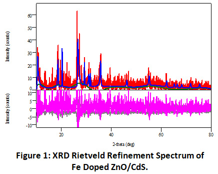 |
Figure 1: XRD Rietveld Refinement Spectrum of Fe Doped ZnO/CdS. Click here to view Figure |
The Rietveld refinement plots of Fe-doped ZnO/CdS nanoparticles was fitted with standard mathematical functions.17,18 The structural analysis is a good agreement with pure hexagonal wurtzite ZnO and CdS phase. From the table it was seen that with increase of doping concentration due to the stress the lattice parameter decreases.
Table 1: Angular Correction.
|
Data name |
a(A) |
b(A) |
c(A) |
alpha(deg) |
Beta (deg) |
gamma(deg) |
|
Fe doped ZnO/CdS |
3.29 (6) |
3.294(6) |
5.27 (11) |
90.000000 |
90.000000 |
120.000000 |
The Rietveld refinement analysis gives information about the variation of lattice constant. The lattice parameter are shown in table. The c/a parameter value is 1.603 which is a close-packed hexagonal structure.
TEM Result Analysis
Composite nanoparticles variation was confirmed by TEM analysis (Model- JEM-100 CX II, Jeol) of the Fe doped ZnO/CdS nanoparticles. It is clear that there was the formation of the distinct, disaggregated and uniform size i.e. monodispersed nanoparticles. Fe doped ZnO/CdS nanostructure showed that the sample had an average diameter of 40 nm with mix spherical and hexagonal shape.
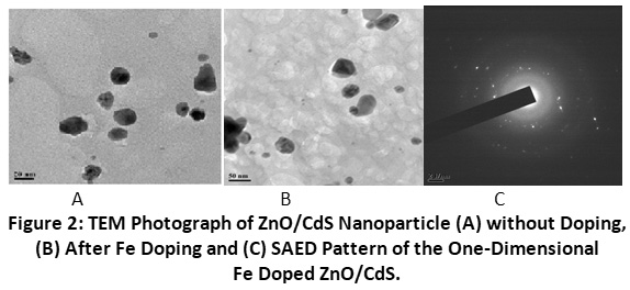 |
Figure 2: TEM Photograph of ZnO/CdS Nanoparticle (A) without Doping, (B) After Fe Doping and (C) SAED Pattern of the One-Dimensional Fe Doped ZnO/CdS. Click here to view Figure |
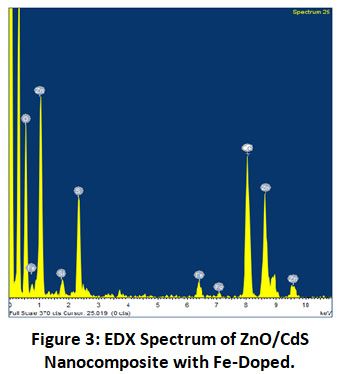 |
Figure 3: EDX Spectrum of ZnO/CdS Nanocomposite with Fe-doped. Click here to view Figure |
Table 2: Elemental Analysis of ZnO/CdS Nanocomposite with Fe-doped.
|
cement |
line |
K factor |
Weight% |
Weight% sigma |
Atomic % |
|
O |
K- SERIES |
1.353 |
9.02 |
1.08 |
33.06 |
|
Si |
K- SERIES |
1.000 |
1.47 |
0.35 |
2.06 |
|
S |
K- SERIES |
1.069 |
9.86 |
0.94 |
6.07 |
|
Fe |
K- SERIES |
2.363 |
4.87 |
0.83 |
3.42 |
|
Zn |
K- SERIES |
3.907 |
48.07 |
1.70 |
38.73 |
|
Cd |
K- SERIES |
2.987 |
26.71 |
0.10 |
16.66 |
|
Total |
|
|
100 |
|
|
Data analysis from the graph and table it is clear that the sample contains S, Fe, Zn and Cd and which is identified only the target elements and found without any impurities. From the above studies it is found that doping of Fe plays vital role for size and properties of the composite nanostructure.
SEM Result Analysis
The morphology of both the dopped and un-dopped with Fe in composite ZnO/CdS NPs were studied by SEM (JSM-6360) (JEOL) Electron microscope. As shown in fig. 4A, the SEM analysis confirmed the size range of 22- 40nm, a clear indication of the formation of ZnO/CdS nanoparticles. Fig 4 (A). shows the formation of NPs that are self-aggregated.
Fig. 4(B) shows the random distribution of ZnO/CdS NPs with 3.0 wt% Fe-doping. The average size found in between 37 to 42 nm. From the SEM analysis it is found that doping concentration changes the particle size.
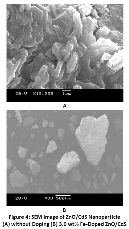 |
Figure 4: SEM Image of ZnO/CdS Nanoparticle (A) without Doping (B) 3.0 wt% Fe-doped ZnO/CdS. Click here to view Figure |
UV Spectrometric Analysis
The UV-Vis spectrum was recorded in the range 300 to 800 nm and shown in Figure 5. For the prepared material ZnO/CdS-doped with Fe, the optical bandgap value found to be 3.83-4.1 eV. It was also found higher doping decreases bandgap. Table shows the variation. The change of lattice mismatch caused by incorporation of Fe2+ the presence of ZnO/CdS matrix may be the possible reason for the variation of band gap.19,20
The absorption spectrum shows four characteristic bands at 360, 406, 465 and 670 nm are due to the transitions from band to band. The position of bands identifies the characteristic of Fe2+doping.
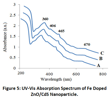 |
Figure 5: UV-Vis Absorption Spectrum of Fe Doped ZnO/CdS Nanoparticle. Click here to view Figure |
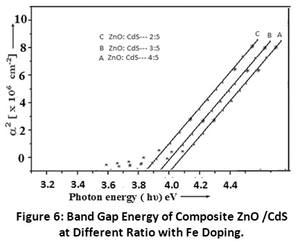 |
Figure 6: Band Gap Energy of Composite ZnO /CdS at Different Ratio with Fe Doping. Click here to view Figure |
From the absorption spectra the possible transition, (αh?)2 versus h? were plotted and calculated the bandgap by extrapolating the straight-line. It is found that band gap is shifted and from this shifting data using established optical model from equation 1 band gap is calculated
Egn = [ E2gb+2?2Egb(π/R)2 / m*]1/2--(1)
Where m* is the average effective mass of the composite, Egb is average bulk band gap and Egn is the estimated band gap energy. With the change of bandgap energy the particle size were estimated with Hyper Band Model(HBM) .It is found that with increase doping concentration of Fe2+ the bandgap increases the particle size decreases according to HBM. The particle size was found in the range 4-9 nm.21,25
Antimicrobial Assay
Antibacterial activity of the Fe doped ZnO/CdS composite nanoparticles was carried out in waterborne pathogenic bacteria species. The antibacterial activity was measured by zone inhibition area by high resolution travelling microscope. Tetracyclin of 1mg/ml, concentration was used as a control antibacterial agent. The results are shown in the Table-2depict that Fe doped ZnO/CdS nanoparticles are efficiently giving zone inhibition.26
Table 3: Antibacterial Efficacy of Fe Doped ZnO/CdS Nanoparticles with Standard Antibiotic Bacterial Strains.
|
Bacterial Strains |
Zone of inhibition (diameter in nm) |
Tetracyclin(1mgml-1) |
||
|
Fe doped ZnO/CdS nanoparticles |
100 μl |
|||
|
50 μl |
80 μl |
100 μl |
||
|
E. coli |
6±0.12 |
10±0.13 |
18±0.11 |
35±0.11 |
|
K.pneumonia |
9±0.09 |
11±0.18 |
20±0.13 |
32±0.12 |
|
P.aeruginosa |
3±0.13 |
8±0.16 |
17±0.09 |
31±0.11 |
|
S. aureus |
6±0.09 |
12±0.11 |
20±0.09 |
34±0.13 |
Conclusions
Fe doped ZnO/CdS nanocomposite were successfully synthesized by chemical method. The prepared sample is composed of mix phases. Due to mismatch of radii of Fe2+ and Zn2+/Cd2+ ions, diffraction peaks shift in the main characteristic peaks of XRD pattern. Optical band gap was calculated for different Fe and ZnO/CdS compositions and modification of band gap from 3.83 to 4.1 eV was observed. Different transitions were indicated by the four optical spectrum peaks. The results are improved for water purification methods including removal of the contaminants from water and destruction or immobilization of toxic compounds and pathogens. The size of the nanoparticles are obtained in different range because shape plays an important role and we have considered only spherical shape from TEM result and also depends on interface.
Acknowledgement
The authors would like to acknowledge NIT Nagaland for providing XRD facilities. We would also like to acknowledge DBT star college scheme and ASTEC for providing financial support through major projects.
Funding Sources
This work is part of financial support of DBT Star college Scheme File no BT/HRD/11/01/2020 and ASTEC file no ASTEC/S&T/192(155)/12-13/3932.
Conflict of Interest
The author(s) declares no conflict of interest.
References
- Irimpan L, Nampoori V.P. N, Radhakrishnan P. Effect of annealing on the spectral and nonlinear optical characteristics of thin films of nano-ZnO. J. Appl. Phys. 2008; 103,094914-094918.
CrossRef - Nayak J, Sahu S. N, Kasuya J, Nozaki S. Synthesis, characterization and application for photocatalytic degradation of 3, 4-dihydroxy benzoic acid. Appl. Surf. Sci.2008; 254(22), 7215-7218.
CrossRef - Yuan, Chen L. Li. F, Chen Y. Nanostructured hybrid ZnO@CdS nanowalls grown in situ for inverted polymer solar cells. J. Mater. Chem. C. 2014;2,1018-1027.
CrossRef - Rao G.T, Babu B, Stella R.J, Manjari V.P, Reddy C.V, Jaesool S, Ravikumar R.V.S.S.N. Structural, optical, and luminescence properties of Cu2+-doped Ca-Li hydroxyapatite nanopowders prepared by mechanochemical synthesis. J Mol Struct. 2015;1081 ,254.
- Brayner R, Ferrari-Iliou, Brivois R , Djediat N , Benedetti S , Fievet M.F. Toxicological Impact Studies Based on Escherichia coli Bacteria in Ultrafine ZnO Nanoparticles Colloidal Medium. Nano Lett. 2016;6, 886.
CrossRef - Thill A, Zeyons O, Spalla O, Chauvat F, Rose J, Auffan M, et al. Cytotoxicity of CeO2 nanoparticles for Escherichia coli. Physico-chemical insight of the cytotoxicity mechanism. Environ. Sci. Technol. 2006;40(19),6151-6156.
CrossRef - Munawar Tauseef, Mukhtar Faisal , Nadeem Shahid Muhammad , Mahmood Khalid , Hussain Altaf , Ali Adnan , Arshad M.I., Nabi M.Ajaz un , Iqbal Faisal. Structural, optical, electrical, and morphological studies of rGO anchored direct dual-Z-scheme ZnO-Sm2O3–Y2O3 heterostructure nanocomposite: An efficient photocatalyst under sunlight. Solid State Sciences.2020;106, 106307.
CrossRef - Munawar Tauseef, Mukhtar Faisal , Nadeem Shahid Muhammad , Mahmood Khalid , Hussain Altaf , Ali Adnan , Arshad M.I., Nabi M.Ajaz un , Iqbal Faisal. Sunlight-induced photocatalytic degradation of various dyes and bacterial inactivation using CuO–MgO–ZnO nanocomposite. Environmental Science and Pollution Research.2021;28, 42243–42260.
CrossRef - Munawar Tauseef, Rehman Muhammad Naveed ur, Nadeem Muhammad Shahid, Mukhtar Faisal, Manzoor Sumaira, Ashiq Muhammad Naeem, Iqbal Faisal. Facile synthesis of Cr-Co co-doped CdO nanowires for photocatalytic, antimicrobial, and supercapacitor applications. Journal of Alloys and Compounds.2021;885. 160885.
CrossRef - Deep A, Kumar P, Narasimhan B, Lim S.M, Ramasamy K, Mishra R.K, Mani V. Azetidinone Derivatives: Synthesis, antimi-crobial, anticancer evaluation and QSAR studies. Acta Polon. Pharm. Drug. Res. 2016; 73, 65-78.
- Keri R.S, Hosamani K.M, Reddy H.S, Shingalapur, R.V. Synthesis, in-vitro antimicrobial and cytotoxic studies of novel azet-idinone derivatives. Arch. Pharm. Chem. Life Sci. 2010 ;343, 237-247.
CrossRef - Rawat D, Nair D. Extended-spectrum β-lactamases in Gram Neg-ative Bacteria. J. Glob. Infect. Dis. 2010; 2(3), 263-274.
CrossRef - Malanovic N, Lohner, K. Antimicrobial Peptides Targeting Gram-Positive Bacteria. Pharmaceuticals (Basel), 2016; 9(3), 59.
CrossRef - Reddy K. M, Feris K, Bell J, Wingett, D .G, Hanley C, Punnoose A. Selective toxicity of zinc oxide nanoparticles to prokaryotic and eukaryotic systems. Appl. Phys. lett. 2007; 90,2139021-2139023.
CrossRef - Perez C, Paul M, Bazeraue P. Antibiotic assay by agarwell diffusion method. Acta Biol Med Exp. 1990; 15, 113-115.
- Ansari, S. A, Nisar A, Fatma B, Khan W, Naqvi A.H. Investigation on structural, optical and dielectric properties of Co doped ZnO nanoparticles synthesized by gel-combustion route. Mater. Sci. Eng. B. 2012; 177(5),428-435.
CrossRef - Zhang C, Huang Z, Liao X, Yin G. Gu. J. Preparation and enhanced ferromagnetic, semi-conductive, and optical properties of Co-doped ZnO rod arrays. J Coat. Technol. Res. 2012; 9(5),621-628.
CrossRef - Sato K, Katayama K, Yoshida H. Material Design for Transparent Ferromagnets with ZnO-Based Magnetic Semiconductors. Jpn. J. Appl. Phys. 2000; 39, L555-L558.
CrossRef - Mohapatra J, Misra D. K, Misra D, Perumal A, Medicherla V. R. R, Phase D. M, et al. Room temperature ferromagnetism in Co doped ZnO within an optimal doping level of 5%. Mater. Res. Bull. 2012;47,1417-1422.
CrossRef - Caglar Y. Sol–gel derived nanostructure undoped and cobalt doped ZnO: Structural, optical and electrical studies. J. Alloy Compd. 2013; 560,181-188.
CrossRef - Barman J, Borah J. P. Sarma K. C. Optical properties of chemically prepared CdS quantum dots in polyvinyl alcohol. Int. J. Mod. Phys. B .2009;23(4), 545.
CrossRef - Barman J, Sarma K. C, Sarma M. Structural and optical studies of chemically prepared nanocrystalline thin films. Indian J. Pure Appl. Phys.2008; 46,394.
- Guinier A. X-Ray Diffraction, Freeman, San Francisco, CA, USA, 1963.
- Pankove J. I. Optical Processes in Semiconductors (Prentice-Hall Inc, Englewood Cliffs, New Jersey, 1971).
- Barman J, Borah J. P, Sarma K. C. Synthesis and characterization of CdS nanoparticles by chemical growth technique”, Journal of Optoelectronics and Advanced Material Rapid Communication.2008;2(12),770.
- Ramyajuliet Madhavan, Priyanka Beulah Gnana raj Gnana, Tresin Pious Soris, Doss Asirvatham, Mohan Veerabahu Ramasamy.Biogenic synthesis of copper nanoparticles using aquatic pteridophyte Marsilea quadrifolia Linn. rhizome and its antibacterial activity.Int. J. Nano Dimens.2020; 11 (4),337-345.


