Bioaccumulation of sodium, potassium, calcium and magnesium in Rohu, Labeo rohita (Ham.) fry
Anusaya Mallick1 * , B.C. Mohapatra1 and N. Sarangni1
1
Central Institute of Freshwater Aquaculture,
Indian Council of Agricultural Research,
Bhubaneswar,
751 002
India
http://dx.doi.org/10.12944/CWE.5.1.17
The bioaccumulation of alkali and alkaline earth metals such as Na, K, Ca and Mg were studied in the fry of rohu, Labeo rohita (Ham.), a species of freshwater aquaculture importance in India. The experiments were conducted in laboratory conditions exposing the rohu eggs up to fry stage in one experiment; and fry to advanced fry stage in other experiment, to different concentrations of the salts. Sodium chloride (0.15, 1.5, 15, and 150 mg/l), potassium chloride (0.015, 0.15, and 1.5 mg/l), magnesium chloride (0.15, 1.5, 15, and 150 mg/l), and calcium chloride (0.15, 1.5, 15, and 150 mg/l) were used as test salts. The tests were conducted in 20-litre capacity glass aquaria stocked with 800 eggs up to fry stage; and in the second experiment with 60 numbers of fry up to advanced fry stage. Control animals were reared in the laboratory without addition of salts. The experiments were conducted in triplicate and each one continued for one-month duration. Normal rearing practices were followed and 50 per cent test solutions from each aquarium were replaced with fresh ones in every week. The bioaccumulations in wet weight basis in whole tissue of fry reared from eggs and in advanced fry exposed to respective salts at the end of the experiment were 100-572 and 91-147 ppm for sodium; 71-104 and 13-22 ppm for potassium; 16-32 and 24-48 ppm for calcium; and 210-660 and 150-300 ppm for magnesium respectively. In control the bioaccumulations for fry reared from eggs, and in advanced fry were 150-212 and 371-412 ppm for sodium; 85-99 and 80-85 ppm for potassium; 18-22 and 5.5-7.8 ppm for calcium; and 250-260 and 15 ppm for magnesium respectively. No definite relationship could be established between the alkali and alkaline earth metals concentration in aquatic environment and bioaccumulation in fish tissue, except sodium and calcium in fry stage.
Copy the following to cite this article:
Mallick A, Mohopatra B. C, Sarangi N. Bioaccumulation of sodium, potassium, calcium and magnesium in Rohu, Labeo rohita (Ham.) fry. Curr World Environ 2010;5 (1):111-116 DOI:http://dx.doi.org/10.12944/CWE.5.1.17
Copy the following to cite this URL:
Mallick A, Mohopatra B. C, Sarangi N. Bioaccumulation of sodium, potassium, calcium and magnesium in Rohu, Labeo rohita (Ham.) fry. Curr World Environ 2010;5 (1):111-116. Available from: http://www.cwejournal.org/?p=1118
Download article (pdf) Citation Manager Publish History
Introduction
The dissolved solids of water mass influence the chemical density of the environment; abundance and composition of the biotic community.¹ The alkaline and alkali earth metals are considered important in aquaculture ecosystem, but their amount if excess is harmful to the aquatic animals. Alkaline metals such as Li, Na, and K, and alkali-earth metal such as Be, Mg, Ca, Sr, Ba and Ra have an approximately equal toxicity for fish.2 Magnesium, calcium and potassium are more toxic to fish than sodium salts.2-3
It is well documented that metals and organic compounds can be accumulated by aquatic biota.4 Bioaccumulation measurements refer to studies or methods monitoring the uptake and retention of these elements in organs and or tissues of organisms, such as fish.5 This can only take place if the rate of uptake by the organism exceeds the rate of elimination.6 There are five potential routes for an element to enter the fish: via., food, non-food particles, gills, oral consumption of water and the skin. Once absorbed, it is transported by the blood to either a strong point (i.e., bone) or to the liver for transformation and/ or storage. According to Heath (1991),7 if the pollutant is transformed by the liver, it may be stored there or excreted in the bile or passed back into the blood for possible excretion by the gills or kidneys, or stored in fat, which is an extra-hepatic tissue. Therefore, the concentration found in different tissues after environmental exposure, for a specific time, depend on several dynamic processes all taking places concurrently.
Material and Methods
Test Container
Glass jar tanks of 20 l capacity were used as test containers in the study. Before use, the jars were cleaned with laboratory detergents, then with 100% acetone and tap water. After each test, the containers were washed appropriately with acid to remove metals and bases; and detergents to remove organic compounds. Each of the test containers was provided with facilities of continuous aeration and covered with velon screen netting to prevent animals from jumping out.
Test Concentration
While designing, minimum of three exposure (treatment) concentrations of a test substance and one control are required to conduct bioassay test and chronic exposure studies. In the present study the test organisms were exposed to a wide range of concentrations selected in logarithmic scale i.e., sodium chloride (0.15, 1.5, 15, and 150 mg/l), potassium chloride (0.015, 0.15, and 1.5 mg/l), magnesium chloride (0.15, 1.5, 15, and 150 mg/l), and calcium chloride (0.15, 1.5, 15, and 150 mg/l). The range of concentrations selected was expected to produce both observed and no-effects.
Test Solutions
The test solutions were prepared by dissolving the calculated amount of salts in the dilution water of the experimental aquaria.
Test Procedure
Two experiments were conducted to evaluate the effects of calcium, magnesium, sodium and potassium on Indian major carp, rohu (Labeo rohita, Hamilton) egg and fry. The experiments were conducted in laboratory conditions exposing the rohu eggs up to fry stage in one experiment; and fry to advanced fry stage in the other experiment, to different concentrations of sodium, potassium, calcium and magnesium salts. The tests were conducted in 20-litre capacity glass aquaria stocked with 800 eggs up to fry stage; and in the second experiment with 60 numbers of fry up to advanced fry stage. Control specimens were reared in the aquaria without addition of salts. Each concentration was tried in triplicate and the test was continued for one-month duration. Normal rearing practices were followed and 50 per cent test media was replaced with fresh ones in every week. During the experimentation period various observations were made in a specific manner and schedule in order to determine the various levels of accumulation of the test elements in fish.
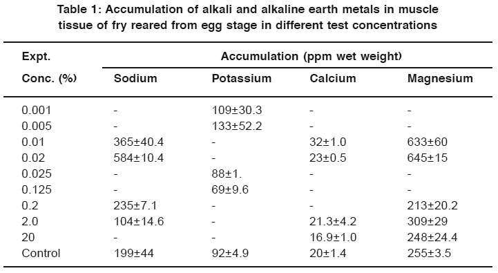 |
Table 1: Accumulation of alkali and alkaline earth metals in muscle tissue of fry reared from egg stage in different test concentrations Click here to view table |
Water Quality Management
Physico-chemical parameters such as temperature, dissolved oxygen, free carbon dioxide, pH, total hardness, total alkalinity, ammonia, nitrite, nitrate, phosphorus and dissolved organic matter of the test media were measured in every week following the standard laboratory methods prescribed in APHA (1998)8. Except pH and temperature all the other parameters were expressed in ppm.
Tissue Collection
The fry (10-16 mm length and 0.006-0.017 g weight) and advanced fry (39-65 mm length and 0.373-1.864 g weight) were harvested from the experiment after one month of exposure to different concentrations of salt solutions in each experiment. From each concentration i.e., each aquaria, fishes were selected randomly for required quantity of samples needed for bioaccumulation estimation.
Digestion and Estimation
The wet weight of fish was estimated from each concentration and one gram tissue (whole body after removal of gut content) was taken and placed into digestion flasks and predetermined amount of concentrated nitric acid was added to the sample. The flasks were kept in the digestion chamber at 120°C until the vapor and the acid fluids inside the flask turned clear. After completion of the digestion process, the samples were cooled and filtered using Whatman filter paper No 42, and the final volume was made up to 25 ml with distilled water. The samples were kept in airtight amber colored glass bottles for determination of the metal concentrations. The concentration of metals was determined using Systronics Flame Photometer (Model 128). Initially the Flame Photometer was calibrated using standard concentrations of sodium, potassium and calcium metal solution, and then reading was taken for samples. Magnesium was estimated by EDTA titration methods.8
All the data recorded from the experiments were tabulated and statistically analyzed.
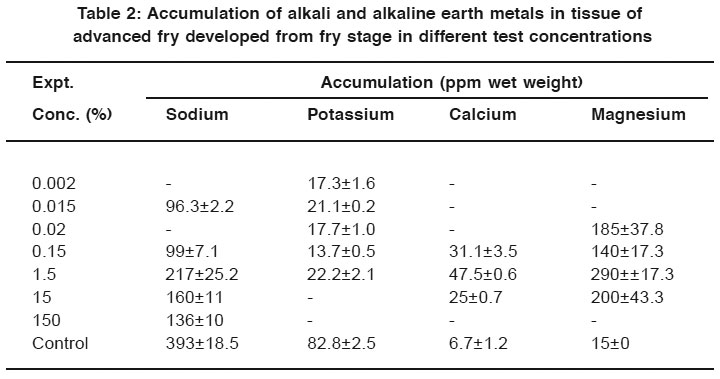 |
Table 2: Accumulation of alkali and alkaline earth metals in tissue of advanced fry developed from fry stage in different test concentrations Click here to view table |
Results and Discussion
The bioaccumulation of sodium, potassium, calcium and magnesium in muscle tissue of the fry and advanced fry in two respective experiments of one month duration is presented in tables 1 and 2.
Experiment 1: Egg to Fry
The sodium bioaccumulation in the tissue of the fry (104±14.6 to 584±10.4) was estimated lower in higher salt concentrations (Fig. 1). Accumulation of the magnesium was not reflected proportionality in the tissue while exposed to higher concentrations of the test salts. Potassium accumulated in fry up to test concentration of 0.005 ppm and then decreased with increase of the salt concentration in the media. Accumulation of calcium (32±1.0 to 17± 1.0) (Table 1 and Fig. 1) decreased from lower concentration to the higher concentration of exposure in case of egg developed to fry.
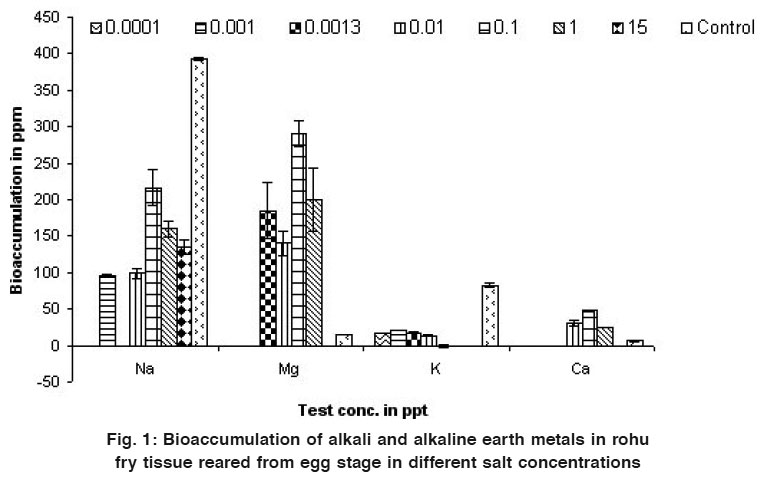 |
Figure 1: Bioaccumulation of alkali and alkaline earth metals in rohu fry tissue reared from egg stage in different salt concentrations Click here to view figure |
In general, at the end of the experiment, bioaccumulations in wet weight basis in whole tissue of fry reared from eggs exposed to respective salts were 100-572 ppm for sodium; 71-104 ppm for potassium; 16-32 ppm for calcium; and 210-660 ppm for magnesium. In control the bioaccumulation in fry reared from eggs was 150-212 ppm for sodium; 85-99 ppm for potassium; 18-22 ppm for calcium; and 250-260 ppm for magnesium.
Experiment 2: Fry to Advanced Fry
The bioaccumulation of sodium in fish tissue varied in different test concentrations and no definite relation was established between them. Bioaccumulation of calcium was increased up to 47.5±0.6 while rohu seed was exposed to the test concentration of 0.15g/l and then after decreased with the increase of concentration. Magnesium and potassium did not follow any trend in their accumulation in muscle tissue (Table 2 and Fig. 2).
The bioaccumulations in wet weight basis in whole tissue of advanced fry exposed to respective salts at the end of the experiment were 91-147ppm for sodium; 13-22 ppm for potassium; 24-48 ppm for calcium; and 150-300 ppm for magnesium. In control the bioaccumulation was 371-412 ppm for sodium; 80-85 ppm for potassium; 5.5-7.8 ppm for calcium; and 15 ppm for magnesium.
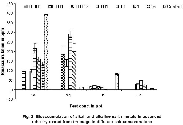 |
Figure 2: Bioaccumulation of alkali and alkaline earth metals in advanced rohu fry reared from fry stage in different salt concentrations Click here to view figure |
Physico-Chemical Properties of Test Solution
The water samples from different experimental aquaria had dissolved oxygen (DO) 4.0-7.6 mg/l, pH 6.5-7.6, total alkalinity 118-240 mg/ l, total hardness 60-700 mg/l, conductivity 0.278-6.34 mili mho/ cm, dissolved carbon dioxide 5-12 mg/l and chloride 40-4700 mg/l.
Metal distribution and accumulation in different tissues of fish varies depending on the sources, uptake, diet and or water borne exposure.9 During early life, the yolk sac stage of fish is considered the most sensitive one, followed by the embryonic stage prior to completion of gastrulation.10 Calcium deficiency includes swelling and poor hatchability of eggs and slow development; lack of resistance and low survival of sac fry. Higher calcium concentration protects the fry from ammonia and metal ecotoxicosis. Magnesium is also essential in skeletal tissue metabolism and neuromuscular transmission.11 Freshwater fish derive magnesium ions by active uptake from the environment or from dietary sources. Fish eggs contain a significant amount of magnesium, and thought that some of it is associated with the yolk.12 Therefore, the yolk may serve as a magnesium source for the developing embryo during the early embryonic stage. In adult tilapia (Oreochromis mossambicus, Peters) a higher magnesium concentration in the ambient waters resulted in hypermagnesaemia and hypocalcaemia.13-14 In the present experiment, all the metals were bioaccumulated more than the control in the fish tissue. The bioaccumulation was seen up to certain level and then after reduced with the increase of their salts in the aquatic environment.
Eggs were considered dead when parts of its content turned opaque and white, or when seen with structural damages, mainly vertebral deformities.15
In the present experiment, the sensitivity of rohu larvae soon after hatching was higher than the control which might be linked to the primary effect of sodium, potassium, calcium and magnesium on embryonic period or early larvae stages, and also to thechemical stress during hatching process. Early life stages of the teleosts are known to be the very sensitive parts of the life. The sensitivity first of all depends on the developmental stages and time of exposure.16-17 physico-chemical characteristic of water, toxicant concentration and fish species.18 In the present experiment the test specimen was exposed to the salts from the very beginning of egg stage to the advanced fry stage. The embryo disintegrated during cleavage and blastula stages when exposed to higher concentration of the salts. In some cases the larvae also deformed when exposed to the higher concentrations of salts.
When fish are exposed to elevated levels of metals in the aquatic environment, they can absorb the bioavailable ones directly from the environment via., gills and skin, or through the ingestion of contaminated water and food. Metals in the fish are then transported by the bloodstream, which brings it into contact with the various organs and tissues.19 Fish can regulate metal concentrations to a certain extent. The ability of each tissue to either regulate or accumulate metals can be directly related to the amount of the metal accumulated in the specific tissue. Furthermore, physiological differences and the position of each tissue in the fish can also influence the bioaccumulation of a particular metal.20
Acknowledgements
We express our sincere thanks to the Director, Central Institute of Freshwater Aquaculture, Bhubaneswar, Orissa, India for providing facilities and allowing the first author to conduct her post- graduate research work.
References
- Jhingran V.G., Fish and Fisheries of India. Hindustan Publ. Co. (India), Delhi (1998).
- Metelev V.V., Kanaev A.I., and Dzasokhova N.G., Water Toxicity. Amerind Publ. Co. Pvt. Ltd., New Delhi (1983).
- Mohapatra, B.C., D. Sc. Thesis, Berhampur University, Orissa (1999).
- USEPA (United States Environmental Protection Agency) Office of Health and Environmental Assessment, US Environmental Protection Agency, Cincinnati, Ohio (1991).
- Roux D., Eastern Transvaal. M.Sc. Thesis, Rand Afr. Univ., South Africa (1994).
- Spacie and Hamelink J.L., Hemisphere Publishing Corporation, New York, USA. (1985) 124-163.
- Heath A.G., Lewis Publishers, Boca Raton, Florida, USA. 359 (1991).
- APHA, American Public Health Association, Water Pollution Central Federation and American Water Works Association, Standard methods for the examination of water and wastewater, 16 th ed. Boyd. Printing Company, Albana, New york. USA (1998).
- Kraal M.H., Kraak M.H.S., DeGroot C.J., and Davids C., Ecotoxicol. Environment. Saft., (1995) 31,179-183.
- Von Westeenhagen H., In W.S. Hoar and D.J. Randall, eds., Fish Physiology, Vol. 11-Academic, London, UK, (1988) pp. 253-346.
- Lall S.P., The Minerals. In: Fish Nutrition (Ed.) J.H. Halver, Academic Press, London-San Diego- California, (1989) 220-252.
- Hayes F. R., Darcy D.A., Sullivan C.M., J. Biol. Chem. (1946) 163: 621-631.
- Wendelaar Bonga S.E., Lowik C. J.M., Van der Meij J.C.A., Gen. Comp. Endocrinol., (1983) 52: 222-232.
- Bijevelds, M.J.C., Flik G., Wendelaar Bonga S.E., Fish Physiology and Biochemistr, (1997) 16: 323-331.
- Eaton J..M., Trans. Am. Fish. Soc. (1974) 103: 121-214.
- McKim J..M., Eaton J.G., and Holcombe G.W., Bulletin of Environmental Contamination and Toxicology, (1978) 19 (5): 608-616.
- Hutchinson T.H., Solbe J. and Kloepper-Sams, P.J.. Chemosphere (1997) 36: 129-142.
- Alabaster, J.S. and Lloyd, R., Butterwotths, London and Boston (1980).
- Vander Putte I. and Part P., Aquatic Toxicol., (1982) 2: 31-45.
- Kotze P.J., Aspects of Water Quality, Mpumalanga. M.Sc. Thesis,Rand Afr. Univ., South Africa (1997).


