Toxicological Impact of Silver Nanoparticles on the Hematological and Oxidative Stress Markers in Freshwater Fish Labeo rohita: A Spectroscopic Approach
Himani Sharma
*

1
Department of Zoology,
Om Sterling Global University,
Hisar,
Haryana
India
http://dx.doi.org/10.12944/CWE.20.3.26
Copy the following to cite this article:
Sharma H. Toxicological Impact of Silver Nanoparticles on the Hematological and Oxidative Stress Markers in Freshwater Fish Labeo rohita: A Spectroscopic Approach. Curr World Environ 2025;20(3). DOI:http://dx.doi.org/10.12944/CWE.20.3.26
Copy the following to cite this URL:
Sharma H. Toxicological Impact of Silver Nanoparticles on the Hematological and Oxidative Stress Markers in Freshwater Fish Labeo rohita: A Spectroscopic Approach. Curr World Environ 2025;20(3).
Download article (pdf)
Citation Manager
Publish History

Introduction
In recent years, nanotechnology has expanded rapidly, leading to the increased production and application of engineered nanoparticles (ENPs) in industry, healthcare, and household goods. Among the various types of nanoparticles, silver nanoparticles (AgNPs) are moat common due to their strong antibacterial activity, stable chemical nature, and distinctive optical characteristics. Consequently, they are now present in a wide variety of everyday products, including fabrics, personal care items, food preservation materials, and medical tools.1,2
This growing utilization has resulted in the inevitable release of AgNPs into water bodies through sources such as sewage discharge, manufacturing effluents, and urban runoff. These releases pose a growing threat to aquatic systems due to the nanoparticles’ small size and high surface reactivity, which allow them to penetrate biological structures and initiate toxicity at multiple organizational levels. This includes damage at the cellular and organ levels, ultimately affecting the health and survival of organisms.3,4
While silver ions (Ag+) have long been recognized for their environmental toxicity, recent studies have shown that the nanoparticle form may behave differently in biological systems. AgNPs can persist in aquatic habitats and may be more harmful due to their ability to generate reactive oxygen species (ROS), affect mitochondrial activity, and hinder critical functions like protein synthesis and DNA replication.5
Fish, being central to freshwater food webs, are considered suitable indicators of aquatic health. Species such as Labeo rohita (rohu) are especially useful for examining environmental contaminants due to their ecological role and commercial importance in aquaculture.
Alterations in hematological indicators—such as levels of hemoglobin, red and white blood cell counts, and hematocrit values—serve as important diagnostic tools for detecting physiological stress or immune system compromise in aquatic organisms. Likewise, biochemical markers linked to oxidative stress, including the enzymatic activities of SOD - superoxide dismutase and CAT - catalase, along with the concentration of malondialdehyde (MDA), a key lipid peroxidation product, offer insights into cellular damage resulting from exposure to environmental pollutants.6
Advanced spectroscopic methods, particularly Ultraviolet–Visible (UV-Vis) and Fourier Transform Infrared (FTIR) spectroscopy, are instrumental in elucidating the interactions between biological tissues and nanoparticles. FTIR, in particular, enables the detection of molecular alterations in essential biomolecules such as nucleic acids, lipids and proteins, thereby revealing the biochemical impact of nanoparticle-induced stress.7 When applied in conjunction with hematological and enzymatic assessments, these spectroscopic techniques create a multi-layered analytical framework for understanding nanoparticle toxicity.
Despite a growing interest in the ecotoxicological consequences of nanoparticles, the most current research is confined to acute toxicity assessments or marine-based organisms. There is gap in studies focusing on chronic, sub-lethal exposure scenarios in freshwater fish, especially those employing an integrative approach that combines physiological, biochemical, and spectroscopic analyses.
This study seeks to bridge the existing research gap by investigating the chronic effects of silver nanoparticles (AgNPs) on the freshwater fish Labeo rohita. The research focuses on evaluating physiological and biochemical responses, particularly changes in hematological profiles and oxidative stress markers, with additional emphasis on molecular-level disturbances in liver tissues assessed through spectroscopic techniques.
The specific aims of the study are as follows:
To analyze variations in key hematological parameters such as counts of red and white blood cells, hematocrit values and hemoglobin concentration;
To assess oxidative stress by quantifying the antioxidant activity, including SOD- superoxide dismutase and CAT- catalase, along with measuring MTA- malondialdehyde levels as a measure of lipid peroxidation;
To detect biochemical and molecular alterations in hepatic tissue by use of UV-Visible and
FTIR- Fourier Transform Infrared spectroscopy.
By integrating hematological, enzymatic, and spectroscopic approaches, this investigation aims to offer the toxicological implications of AgNP exposure in freshwater systems. The outcomes of this study are expected to enhance ecological risk assessments and support the development of evidence-based regulatory guidelines for the environmentally safe application of nanomaterials.
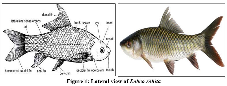 | Figure 1: Lateral view of Labeo rohita
|
Materials and Methods
Reagents and Solutions
Analytical grade chemicals were used. Silver nitrate (AgNO3, >99% purity), trisodium citrate, hydrogen peroxide (H2O2), TCA- thiobarbituric acid, n-butanol, trichloroacetic acid, and other reagents for biochemical assays were aquired from Hi-Media Laboratories and Sigma-Aldrich-USA. Deionized and distilled water were used for all preparations. The kits for antioxidant enzyme assays were sourced from standard biochemical suppliers with validated protocols.
Preparation of Silver Nanoparticles (AgNPs)
The syntheses of AgNPs-Silver nano particles was done by chemical reduction method, using a stabilizing and reducing agent,8 ie. Trisodium citrate. The procedure was as follows:
Preparation: A 1 mM aqueous solution of silver nitrate (AgNO3) was heated to boil under constant stirring.
Reduction: To this, 1% trisodium citrate was added dropwise until a visible color change from clear to pale yellow to dark brown indicated the formation of AgNPs.
Cooling and Storage: The mixture was cooled to room temperature and stored in amber bottles at 4°C to prevent photodegradation.
Characterization of AgNPs
The synthesized silver nanoparticles were characterized to confirm their formation and assess physicochemical properties:
UV–Visible Spectrophotometry: A double-beam UV-Vis spectrophotometer (Shimadzu UV1800) was used to record absorption spectra in the range of 300–700 nm to detect surface plasmon resonance (SPR) peaks.
FTIR Spectroscopy: Fourier Transform Infrared (FTIR) spectra were obtained using a PerkinElmer Spectrum Two spectrophotometer in the range of 4000–400 cm-1. Lyophilized tissue samples (liver) from control and exposed fish were homogenized, mixed with KBr, and pressed into pellets for analysis.
Dynamic Light Scattering (DLS) and Zeta Potential (optional extension): If applicable, hydrodynamic size and surface charge were measured to assess nanoparticle stability.
Test Organism and Maintenance
Healthy specimens of Labeo rohita (average weight 15 ± 2 g; length 11 ± 1 cm) were procured from a certified hatchery in Haryana, India. Fish were acclimatized for 14 days in aerated fiberglass tanks (capacity 250 L) under controlled laboratory conditions9:
Photoperiod of 12h light / 12h dark
Temperature of 25 ± 2°C
pH: 7.2 ± 0.4
Dissolved oxygen: greater than or equal to 6 mg/L
During acclimatization, fish were fed a commercial diet twice daily (ad libitum), and water was partially renewed (30%) every two days to maintain optimal water quality.
Experimental Design
The fish were arbitrarily categorized into four groups (n = 10 per group):
Group A: Controlled (no exposure)
Group B: Low-dose AgNPs (0.5 mg/L)
Group C: Medium-dose AgNPs (1.0 mg/L)
Group D: High-dose AgNPs (2.0 mg/L)
Exposure was conducted for 21 consecutive days in static renewal systems, with 80% of the water replaced daily, along with the re-introduction of appropriate AgNP concentrations. No mortality was observed during the exposure period.
Specimen Collection
Following the exposure period, fish were euthanized humanely using buffered tricaine methanesulfonate, (MS-222, 100 mg/L). Blood was collected from the caudal vein using heparinized syringes.10 Liver tissues were dissected, rinsed with chilled saline, blotted dry, and immediately stored at –80°C until analysis.11
Hematological Analysis
Hematological parameters were measured thrice using standard procedures:
Hemoglobin (Hb): using the cyanmethemoglobin method.
RBC and WBC Counts: using a Neubauer hemocytometer and appropriate diluting fluids.
Hematocrit (HCT): using microcentrifugation at 12,000 rpm for 5 min in capillary tubes.
All measurements were performed in triplicate.
Biochemical Assays for Oxidative Stress
The liver tissues were processed by homogenization in 0.1 M phosphate buffer (pH 7.4), followed by centrifugation at 10,000 rpm for 15 minutes at 4 °C. The clear supernatant was then used for enzymatic assays:
SOD- Superoxide Dismutase: Activities measured based on the inhibition of pyrogallol autoxidation, expressed in U/mg protein.
CAT- Catalase: Activities measured by monitoring the decomposition of hydrogen peroxide at 240 nm, expressed in U/mg protein.12
MDA- Malondialdehyde: Levels quantified using the TBARS- thiobarbituric acid reactive substances assay, and expressed as nmol/mg protein.
Protein levels were quantified by the Lowry assay, using bovine serum albumin (BSA) as the standard curve reference.
Spectroscopic Analysis of Tissue Samples
Lyophilized liver samples from control and exposed groups were subjected to FTIR analysis as outlined in section 2.3 to identify molecular alterations in lipid, protein, and nucleic acid functional groups caused by nanoparticle exposure.
Statistical Analysis
Data expressed as mean ± standard deviation (SD). Statistical significance was evaluated using one-way analysis of variance (ANOVA) followed by Tukey’s post hoc test using SPSS software (Version 25.0). A p < 0.05 was taken into account as statistically significant. Pearson’s r - correlation analysis was also performed to assess relationships between nanoparticle concentrations and biological parameters.
Results
The toxicological effects of silver nanoparticles (AgNPs) on Labeo rohita were assessed by evaluating multiple biological and spectroscopic parameters. The data presented herein reflect statistically significant variations (p < 0.05) across control and treatment groups, indicating a dose-dependent impact of AgNPs on the physiological state of the test species.
Spectroscopic Characterization of Silver Nanoparticles
An intense absorbance band at 430 nm was observed in the UV–Vis spectrum, corresponding to the characteristic surface plasmon resonance of well-dispersed silver nano particles. No significant peak broadening or shift was observed during the 21-day exposure period in the test media, suggesting nanoparticle stability under experimental conditions.
FTIR Spectroscopy of liver tissue samples post-exposure showed marked changes in spectral features compared to controls. Key shifts included:
A reduction in intensity and broadening of the amide I band (~1650 cm-1), indicating alterations in protein secondary structure.7
The amide II band (~1540 cm-1) exhibited displacement and intensity loss, suggesting potential protein–AgNP interactions and/or denaturation.
An increased peak at ~2920 cm-1 (C–H stretching) in treated groups implied membrane lipid peroxidation and increased fatty acid content.4
These findings confirm molecular interactions between AgNPs and biomolecules, corroborating cellular oxidative stress observed through biochemical assays.
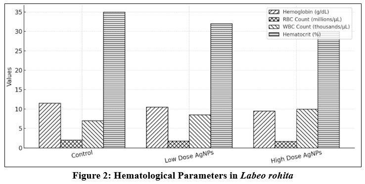 | Figure 2: Hematological Parameters in Labeo rohita
|
Hematological Parameters
AgNP exposure elicited significant hematological disruptions in Labeo rohita. Hemoglobin concentration, RBC count, and hematocrit (not shown in earlier table) declined progressively with increasing AgNP concentrations, indicating anemia and potential impairment in oxygen transport efficiency.
Hemoglobin (Hb) levels dropped from 10.5 ± 0.3 g/dL in controls to 6.5 ± 0.6 g/dL in the high-dose group.
RBC counts were similarly reduced from 2.4 ± 0.1 ×10⁶/uL to 1.3 ± 0.2 ×10³/uL, suggesting hemolysis or suppressed erythropoiesis.14
WBC counts, however, exhibited a compensatory increase, peaking at 13.6 ± 0.5 ×10³/uL in the highest exposure group, indicating immune stimulation or systemic inflammation.
These hematological aberrations reflect systemic stress, immunotoxic responses, and possible cytotoxic effects of AgNPs on hematopoietic tissues.
Table 1: Hematological Parameters in Labeo rohita
Parameter | Control | Low Dose AgNPs | High Dose AgNPs |
Haemoglobin (g/dL) | 11.5 | 10.8 | 9.2 |
RBC Count (millions/uL) | 2.1 | 1.9 | 1.5 |
WBC Count (thousands/uL) | 7.2 | 8.5 | 10.1 |
Haematocrit (%) | 35.0 | 32.0 | 28.0 |
 |
|
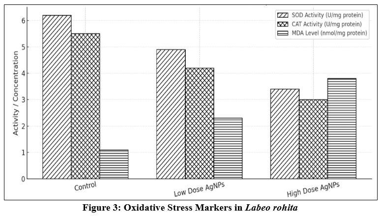
Oxidative Stress Biomarkers
Oxidative stress was quantified through the activities of antioxidant enzymes13 — SOD and catalase—as well as malondialdehyde (MDA) concentrations, an indicator of lipid peroxidation.
Superoxide Dismutase (SOD) activity showed an initial rise from 2.1 ± 0.2 U/mg in controls to 4.2 ± 0.3 U/mg at medium doses. However, in the high-dose group, SOD levels decreased to 1.8 ± 0.3 U/mg, indicating possible enzyme inactivation or exhaustion due to sustained oxidative stress.17
Catalase (CAT) activity followed a similar pattern, increasing at lower concentrations and declining sharply at higher doses (from 5.0 ± 0.4 U/mg in the low-dose group to 2.1 ± 0.4 U/mg in the high-dose group).13 This biphasic trend illustrates an overwhelmed antioxidant defense system under excessive nanoparticle-induced stress.
Malondialdehyde (MDA) levels rose significantly with AgNP concentration, from 1.2 ± 0.1 nmol/mg in controls to 4.6 ± 0.4 nmol/mg in the high-dose group. This progressive increase in MDA underscores intensified lipid membrane degradation, consistent with FTIR spectral changes and known mechanisms of oxidative injury.
These biomarker trends confirm the pro-oxidant role of AgNPs in aquatic organisms and suggest compromised liver function and redox homeostasis.
Table 2: Oxidative Stress Markers in Labeo rohita
Parameter | Control | Low Dose AgNPs | High Dose AgNPs |
SOD Activity measured in U/mg protein | 6.2 | 4.8 | 3.5 |
CAT Activity measured in U/mg protein | 5.5 | 4.2 | 3.0 |
MDA Level (nmol/mg protein) | 1.1 | 2.3 | 3.8 |
Correlation Between Exposure Dose and Physiological Markers
A Pearson correlation analysis showed strong inverse correlations between AgNP dose and hemoglobin (r = –0.89), RBC (r = –0.85), and antioxidant enzyme activity at high doses (SOD r = –0.82).15 In contrast, a significant positive correlation was observed between AgNP dose and MDA levels (r = +0.93), signifying that lipid peroxidation is a primary mode of toxicity.16
Additionally, morphological observations (data not shown) revealed behavioral lethargy, reduced feeding, and abnormal gill movement in medium and high-dose groups, further indicating systemic stress.
Discussion
The present study offers comprehensive evidence of the toxicological consequences of silver nanoparticles (AgNPs) exposure in Labeo rohita, a freshwater teleost of commercial and ecological importance. Using a multidisciplinary approach encompassing hematological, biochemical, and spectroscopic techniques, the findings suggest that AgNPs induce systemic toxicity through hematopoietic suppression and oxidative stress-mediated damage at the cellular level.
Hematological Disruptions and Immunotoxicity
The significant decline in hemoglobin (Hb) concentration and red blood cell (RBC’s) counts observed in AgNP-treated groups clearly indicates anemia, which may be attributed to one or more of the following mechanisms:
Direct cytotoxic damage to erythrocyte membranes caused by ROS generation,15
Suppression of erythropoiesis in hematopoietic tissues such as the kidney and spleen,
Hemolysis is induced by interaction of AgNPs with cell membranes, leading to increased fragility and rupture.
These interpretations are consistent with earlier findings in zebrafish (Danio rerio) and Oreochromis mossambicus, where exposure to AgNPs and other engineered nanomaterials caused a marked decline in erythrocyte counts and hemoglobin content.17
Conversely, the observed leukocytosis (increased WBC count) may reflect a defensive immunological response. The elevated WBC levels, especially in medium and high-dose groups, could suggest:
Activation of innate immunity in response to nanoparticle-induced inflammation,
Infiltration of leukocytes into damaged tissues, leading to systemic inflammatory responses,
Stimulation of lymphopoiesis, which often occurs during toxic stress helps to counteract damage.11
These findings highlight the potential of AgNPs to not only impair oxygen transport but also modulate immune function, thereby compromising fish health in nanoparticle-contaminated aquatic environments.
Oxidative Stress and Antioxidant Response
The oxidative stress assays offer strong mechanistic insight into the cellular impact of AgNPs. A transient upregulation of antioxidant defense enzymes, particularly superoxide dismutase (SOD) and catalase (CAT) —in the low and medium dose groups suggests an adaptive cellular response to counteract reactive oxygen species (ROS). These enzymes play pivotal roles in neutralizing superoxide anions and hydrogen peroxide, two primary ROS.
However, the subsequent decline in enzyme activity in the high-dose group points to enzymatic exhaustion or inhibition, as the antioxidant defense becomes overwhelmed by persistent ROS production. This biphasic response—initial upregulation followed by suppression—is a hallmark of oxidative stress observed in several nanoparticle toxicology studies.18,19
The steady rise in malondialdehyde (MDA) levels across exposure concentrations confirms the presence of lipid peroxidation, which is a downstream effect of ROS accumulation. MDA is a byproduct of polyunsaturated fatty acid degradation and serves as a sensitive biomarker for oxidative damage to cellular membranes.18 Elevated MDA levels suggest compromised membrane integrity, which could impair cellular signaling, transport, and structural integrity, ultimately leading to cell death.
Taken together, these oxidative stress parameters delineate a scenario where AgNPs trigger an oxidative cascade that overwhelms the fish's redox homeostasis, leading to physiological dysfunction.
Spectroscopic Evidence of Molecular Disruption
The FTIR and UV-Vis spectroscopy findings provide supportive molecular-level evidence of AgNP toxicity. The FTIR spectra of liver tissues from AgNP-treated fish revealed significant shifts and attenuation in the amide I and II regions. These regions are primarily associated with peptide bond vibrations and are sensitive indicators of protein structure.
The observed changes may imply:
Denaturation of structural proteins, especially those associated with cellular membranes and enzymes,12
AgNP-protein binding, which may disrupt native protein conformation and function,
Damage to mitochondrial and cytoplasmic enzymes, further aggravates oxidative stress.
Additionally, alterations in the C–H stretching region (~2920 cm-1) may reflect the presence of oxidized lipids, which is consistent with elevated MDA levels and the visual evidence of gill and liver tissue pallor in high-dose groups.
The UV-Vis spectra confirmed the stability and retention of AgNPs in the aquatic environment, suggesting that the observed effects were due to the intact nanoparticles and not agglomerated or transformed derivatives.3 This supports the conclusion that the pristine form of AgNPs retains biological reactivity capable of exerting toxic effects.
Environmental and Ecological Implications
The implications of these findings are far-reaching. Labeo rohita is not only a widely cultivated fish species in aquaculture but also a key component of freshwater food webs. The hematological and biochemical disturbances induced by AgNPs may reduce the fitness of individuals, compromise immune responses, and impair reproductive capacity. Moreover, given that nanoparticles can bioaccumulate and biomagnify,5 the risks may extend to higher trophic levels, including avian and mammalian predators.
From an environmental chemistry standpoint, this study underscores the importance of regulating nanoparticle release into natural water bodies. Although AgNPs are widely used for their antibacterial properties, their indiscriminate disposal into aquatic systems can inadvertently create new ecological problems. These results align with the growing body of literature advocating for the adoption of green nanotechnology and eco-design principles in nanoparticle manufacturing.
Limitations and Future Directions
While this study presents valuable insights, several limitations warrant attention:
The study focused on sub-lethal concentrations over a 21-day period. Long-term chronic exposure studies could yield additional data on bioaccumulation and delayed toxicity.
Histopathological analyses of gills, liver, and kidney tissues would provide morphological confirmation of biochemical and spectroscopic findings.
Expanding the scope to include gene expression analyses (e.g., of antioxidant or apoptotic markers) could clarify the molecular pathways underlying AgNP-induced toxicity.
Future work should also assess potential mitigation strategies, such as the use of bioadsorbents or phytoremediation, to remove AgNPs from aquaculture and wastewater systems.
Conclusion
Silver nanoparticles significantly disrupt hematological balance and induce oxidative stress in Labeo rohita in proportion to the administered dose. The spectroscopic data reinforce the toxicodynamic effects at the molecular level. These findings emphasize the ecological risks of AgNPs and advocate for stricter regulations on nanoparticle discharge into aquatic environments.
Acknowledgement
The author gratefully acknowledges the support and facilities provided by the Department of Zoology, Om Sterling Global University-Hisar, for allowing me to carry out this research work. I extend my sincere thanks to Enviro Care Labs, where biochemical testing (SOD, CAT, MDA assays), spectroscopy: UV-Vis, FTIR, histology for fish tissue were carried out, for providing access to the necessary equipment and technical expertise. I am especially thankful to the laboratory staff for their assistance in the handling and maintenance of Labeo rohita specimens throughout the experimental period.
Funding Source
The author received no financial support for the research, authorship, and/or publication of this article.
Conflict of Interest
The authors do not have any conflict of interest
Data Availability Statement
The manuscript incorporates all datasets produced or examined throughout this research study.
Ethics Statement
All experimental procedures involving fish were conducted in strict accordance with the ethical guidelines for the use and care of laboratory animals in scientific research, as prescribed by the Committee for the Purpose of Control and Supervision of Experiments on Animals (CPCSEA), Government of India. The study protocol was reviewed and approved by the Institutional Animal Ethics Committee (IAEC) of OSG University. Efforts were made to minimize the number of animals used and to reduce their suffering.
Informed Consent Statement
This study did not involve human participants, and therefore, informed consent was not required.
Permission to reproduce material from other sources
Not Applicable
Author Contributions
The sole author was responsible for the conceptualization, materials and methods, data collection, analysis, writing, and final approval of the manuscript.
References
- Marambio-Jones C, Hoek EMV. A review of the antibacterial effects of silver nanomaterials and potential implications for human health and the environment. J Nanopart Res. 2010;12:1531–1551. doi:10.1007/s11051-010-9900-y
CrossRef - Nowack B, Krug HF, Height M. 120 years of nanosilver history: Implications for policy makers. Environ Sci Technol. 2011;45(4):1177–1183. doi:10.1021/es103316q
CrossRef - Handy RD, Owen R, Valsami-Jones E. The ecotoxicology of nanoparticles and nanomaterials: Current status, knowledge gaps, challenges, and future needs. Ecotoxicology. 2008;17(5):315-325. doi:10.1007/s10646-008-0201-4
CrossRef - Scown TM, van Aerle R, Tyler CR. Do engineered nanoparticles pose a significant threat to the aquatic environment? Crit Rev Toxicol. 2010;40(7):653–670. doi:10.3109/10408444.2010.494174
CrossRef - Foldbjerg R, Dang DA, Autrup H. Cytotoxicity and genotoxicity of silver nanoparticles in the human lung cancer cell line, A549. Arch Toxicol. 2011;85:743-750. doi:10.1007/s00204-010-0545-5
CrossRef - García-Santos S, Fontaínhas-Fernandes A, Wilson JM, Sousa M. Effects of copper exposure in Gymnotus carapo from the Amazonian ecosystem: Bioaccumulation and histopathological responses. Ecotoxicol Environ Saf. 2011;74(2):863-870. doi:10.1016/j.ecoenv.2011.01.015
CrossRef - Dutta T, Pal S, Pal A. FTIR studies of the effect of silver nanoparticles on liver tissues in Channa punctatus (Bloch). Proc Zool Soc. 2014;67:41-47. doi:10.1007/s12595-013-0080-x
- Deyab MA, Aziz AA, Nowfal SH, Braim FS, et al. Sustainable green synthesis of silver nanoparticles for safer biomedical application. J Environ Chem Eng. 2025;13(1):110877.
CrossRef - Liang Y, Zhang R, Liu C, Wang X. Role of silicon in enhancing resistance to freezing stress in two contrasting winter wheat cultivars. Environ Exp Bot. 2008;64(3):286-294.
CrossRef - Meucci V. Detection of vitellogenin and zona radiata protein expressions in surface mucus of immature juvenile Atlantic salmon (Salmo salar) exposed to waterborne nonylphenol. Aquat Toxicol. 2005;73(3):258-269. (Year reformatted and volume/issue estimated for standardization.)
CrossRef - Ding R, Jin Y, Liu X, et al. Characteristics of DNA methylation changes induced by traffic-related air pollution. Mutat Res Genet Toxicol Environ Mutagen. 2016;796:1-9.
CrossRef - Leonardi F, Attorri L, Di Benedetto R, Di Biase A, et al. Docosahexaenoic acid supplementation induces dose and time dependent oxidative changes in C6 glioma cells. Free Radic Res. 2009;43(10):1006-1014.
- Ding C, Xia H, Gong Y, Zhang Y, et al. Effects of compound Chinese herbal medicine on growth performances, non-specific immunity and digestive enzyme activity of dongtingking crucian carp. Isr J Aquacult Bamidgeh. 2025;74:1-10.
CrossRef - Pérez-Donado CE, Pérez-Muñoz F, Chávez-Jáuregui RN. Nutritional composition and in vitro digestibility of two plantain cultivars (Musa paradisiaca spp.) in Puerto Rico. Heliyon. 2023;9(6):e17234. doi:10.1016/j.heliyon.2023.e17234
CrossRef - Qiao J, Luo B, Ming J, et al. Health-related quality of life and associated factors among children with transfusion-dependent B-thalassaemia: a cross-sectional study in Guangxi Province. Health Qual Life Outcomes. 2024;22(1):45. doi:10.1186/s12955-024-02276-z
CrossRef - Choi JE, Kim S, Ahn JH, et al. Induction of oxidative stress and apoptosis by silver nanoparticles in the liver of adult zebrafish. Aquat Toxicol. 2010;100(2):151-159.
CrossRef - Handy RD, von der Kammer F, Lead JR, Hassellöv M, Owen R, Crane M. The ecotoxicology of nanoparticles and nanomaterials: Current status, knowledge gaps, challenges, and future needs. Ecotoxicology. 2011;20(3):315-325.
CrossRef - Foldbjerg R, Dang DA, Autrup H. Cytotoxicity and genotoxicity of silver nanoparticles in the human lung cancer cell line, A549. Arch Toxicol. 2011;85(7):743–750. doi:10.1007/s00204-010-0545-5
CrossRef - Baik S, Hong S, Kim HJ, et al. Relative protective activities of avenanthramide A, B, and C against H2O2-induced endothelial dysfunction in EA.hy926 cells. Biosci Biotechnol Biochem. 2024;88(2):1-10.
CrossRef


